Syn. Steinernema anomalae (Kozodoi, 1984) Curran, 1989
Measurements: Length=1845 micrometers (um) (1280-2320), Width=117 um (70-165), esophagus length=168 um (125-209), Tail=32 um (14-40), anterior end to testis flexure=441 um (230-690), Spicule=76 um (63-93)[mean=84 um was reported by Kozodoi, 1984],gubernaculum=53 um (45-63), D=60 (38-127).
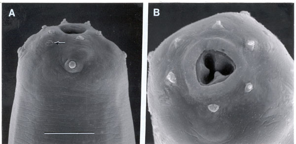
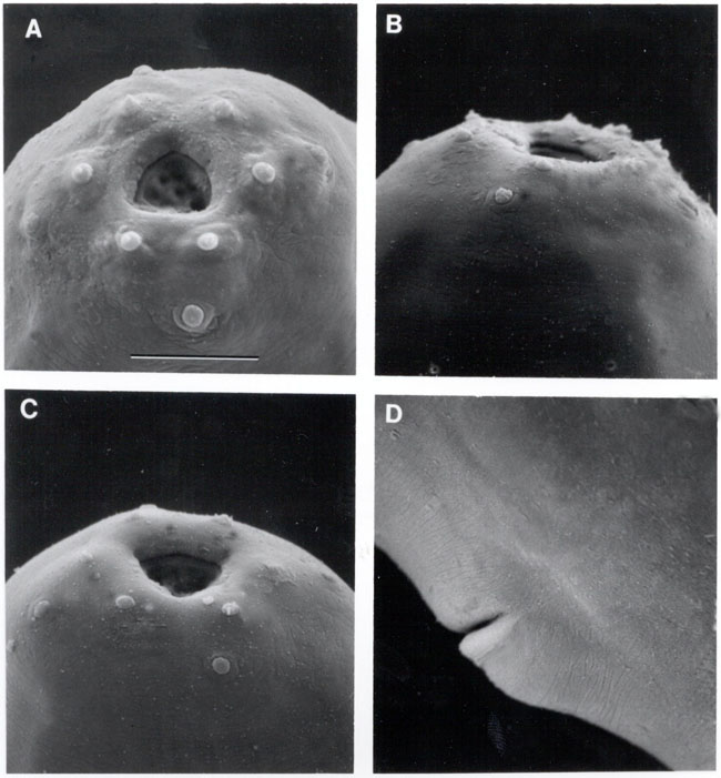
Females: (FemaleSEM) Body with yellowish intestine and colorless gonads. Anterior end broadly rounded with six pointed labial papillae and four rounded cephalic papillae. In stoma, an anterior cuticularized ring present, 1.5 um thick, posterior part similar to that of other steinernematids. Esophagus with prominent metacorpus. Valves in basal bulb present. Excretory duct 3 um wide anteriorly. Anterior gonad in some specimens few eggs were observed in uteri situated mainly in dorsal and lateral sectors of body; posterior gonad having more loops and passing through all sectors of the body. Uterus having two parts: the part near vulva with walls consisting of swollen cells with two-three nuclei, and the part near ovary having thinner walls. In young females these parts of the uterus are divided by a constriction. Tail with rounded terminus, about one fourth of the population having 2-3 um long spike, two such spikes were observed in one female.
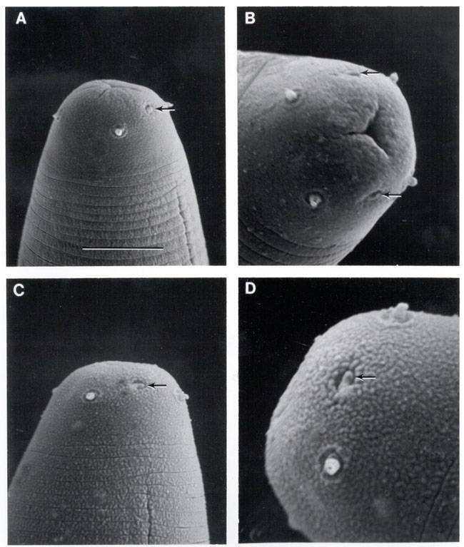
Infective juveniles: (IJSEM) Body long and thick. Lateral field with 8 ridges (9 lines). Anterior end rounded, occasionally with slightly offset cephalic capsule, 12-14 um wide. Tail conical with spherical or irregular border between hypodermal and hyaline parts.
Measurement Length=1217 micrometers (um)(930-1580), Width=37 um (31-44), esophagus length=156 um (132-187),Tail=81 um (65-95, a=33 (25-40), b=7.8 (5.9-9.2), c=15.1 (12.2-17.9),D=63 (53-68).
Features of live S. arenarium. In female: in the postovarial part of the uterus, large moving spermatozoa can be seen. Immobile cells with coarse surface (probably spermatocytes) can be seen close to the constriction in both anterior and posterior branches of young female gonad tubes. In infective juvenile: structures resembling bacterial vesicles can be seen behind the cardia in some juveniles as a transparent sack-like body with numerous folds inside. No separate bacterial cells can be distinguished inside it.
Neotype: A neotype male of S. arenatium (Ailyukliovsky, 1967) has been deposited in the collection of the Zoological Museum of Moscow State University (slide number Ic-396).
References:
Artyukhovsky,
A. K. E. M. Kozodoi, A. P. Reid and S. E. Spiridonov 1997.Redescription
of Steinernema arenarium (Artyukhovsky, 1967) topotype from Central Russia
and a proposal for S. anomalae (Kozodoi, 1984) as a junior synonym. Russian
Journal of Nematology 5:31-37.
Kozodoi, E. M. 1984. A new entomopathogenic nematode Neoaplectana anomali sp. n. (Rhabditida, Steinernematidae) and observations on its biology). Zoologychesky Zhurnal 63:1605-1612.
Nguyen, K. B. and G. C. Smart, Jr. 1993. Scanning electron microscope studies of Steinernema anomali Kozodoi, 1984. Journal of Nematology 25:486-492.