HETERORHABDITIS
TAYSEARAE
Shamseldean, El-Sooud, Abd-Elgawad & Saleh, 1996
Measurement
See Table 1
Description
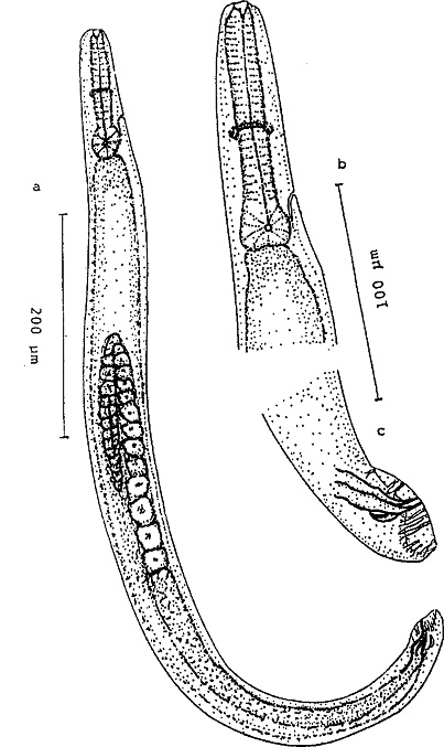
Males (Fig. 1)
Body curved ventrally posteriorly when relaxed, or killed.
Head truncate, or rarely slightly rounded. Six distinct protruding lips surrounding the mouth with 6
papillae on the inner lips Amphids
oval. Cheilorhabdions present as a retractile
ring just below the lips. Pro- and
mesorhabdions reduced, bearing a large tooth near the posterior end of
protorhabdions. The pharynx with fairly
indistinct metacorpus but with a
short isthmus and obvious basal bulb containing fine striations in the valve area. Nerve ring distinct, located at the anterior part of the isthmus
in males and females. Testis single, reflexed, spicules paired,
separated, curved and with pointed tips. Gubernaculum almost half the length of
spicules, relatively broad and
curved. Bursa peloderan with 9 pairs
of genital papillae. From anterior to posterior, pair 1 well anterior, pairs 2
and 3 in a group right anterior to cloaca; pairs 4, 5 and 6 forming a group,
just posterior to cloaca. Pairs 7 and 8, not reaching rim of bursal
membrane. The rest of the genital papillae
are straight and reach the bursal rim.
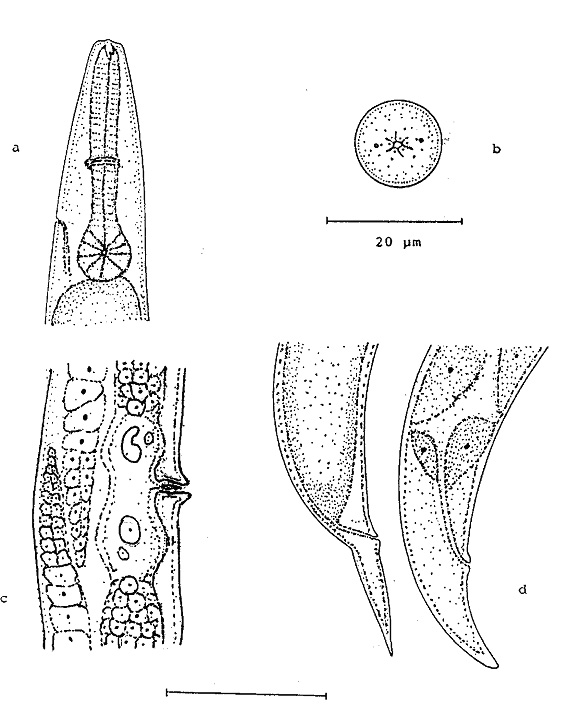
Anterior part is similar to that of male but much larger. Hermaphroditic and amphimictic females with paired, amphidelphic ovaries always with re-flexed portion extending past the vulvar opening. Hermaphroditic females with sperms occurring in the proximal portion of the ovotestis; amphimictic females with sperms in the proximal portion of the oviduct. Vulval region protuberant without copulation plug. Anal swelling is more pronounced in hermaphroditic than amphimictic females. The rectum slightly sclerotized.
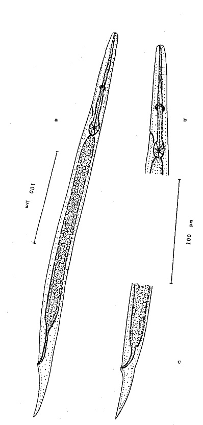
Body length
average of the third stage juveniles (inside second stage
cuticles) are shorter than
that of previously published species. Tail length is too short, cuticle with
fine longitudinal striations. Mouth and anus
closed. Opening of excretory pore located above
level of basal bulb. Nerve ring in the
anterior part of isthmus. The lips of the third stage juveniles contain a faint dorsal tooth. The stoma walls are opened and not collapsed at the base.
The outer second stage cuticle is closely oppressed to the third stage cuticle. The hemizonid distinct. The
ventricular portion of the intestine is devoid of intestinal cells and forms an intestinal pouch which is filled with the symbiotic bacteria. These bacteria occur
in the intestinal lumen, mainly in
the anterior portion.
Diagnostic
characters
This
nematode is characterised by the short body of infective juvenile 418 (332-499)
μm, short tail length 55 (44-70) μm, and high value of E (1.8). Excretory
pore of the male was reported to be anterior to base of esophagus.
Type
host and locality
The nematode was collected from soil in Al-Husane village
in West Nubaria, Behera
governorate, Egypt.
Type material
Holotype: Amphimictic female isolated from the hemocoel of G. mellonella Last
instar larvae (Lepidoptera: Galleridae),
infected by the nematodes through the exposure to the above-mentioned soil
samples. The slide is deposited in the Nematode collection at Faculty of Agriculture, University of Cairo, Egypt.
Distribution
The nematode was found in Egypt (Shamseldean, El-Sooud, Abd-Elgawad & Saleh, 1996).
Reference
Shamseldean, M.M., Abou El-Sooud, A.B., Abd-Elgawad, M.M.
& Saleh, M.M. (1996). Identification
of a new heterorhabditid species from Egypt, Heterorhabditis
taysearae, n. sp.(Rhabditroa: Heterorhabdrndae). Egyptian
Journal of Biological Pest Control 6, 129-138