Steinernema akhursti Qiu, Hu, Zhou,
Mei, Nguyen & Pang, 2005
Description
Male,
first-generation
Body
usually J-shaped when killed by gentle heat. Head rounded, continuous with body
contour. Six labial papillae, two amphids and four cephalic papillae prominent.
Stoma shallow, cheilorhabdions and pro-rhabdions sclerotized, small, sometimes
indistinct. Pharynx muscular with cylindrical procorpus, metacorpus slightly
swollen, isthmus distinct and surrounded by nerve ring, basal bulb prominent.
Excretory pore located anterior to nerve ring, at the base of metacorpus. Gonad
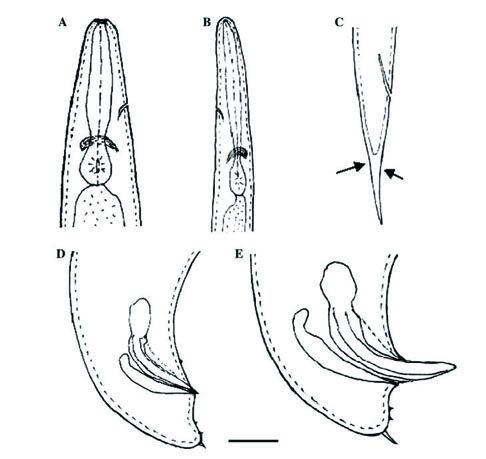
monorchic, reflexed. Spicules paired, brown in color. The length of spicule
head is greater than width, blade moderately curved, dorsal lobe and lateral
lobe prominent, starting from the head and extending to the tip of blade.
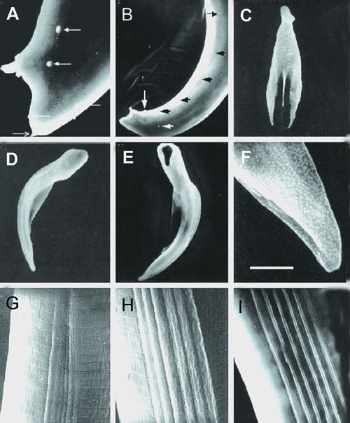
Ventral lobe indistinct. Spicule tip blunt with an aperture on ventral side and
close to the tip (Fig. 1). Velum well developed,
starting from the anterior end of the ventral lobe and ending at one third of
spicule length from the spicule tip. Gubernaculum boat-shaped in lateral view,
tapering gradually anteriorly. Anterior part of the gubernaculum bending
ventrally (Fig. 2). Cuneus long, needle-shaped.
Eleven pairs and one single precloacal genital papilla distributed as in Fig. 1D and E. Tail conoid, usually
concave on ventral side.Tail tip with a prominent mucron.
Similar
to that of the first generation but most morphometrics such as body, spicule
and gubernaculum length smaller. Tail mucron is 1.5 to 2 times as long as that
in the first generation male.
Female, first generation
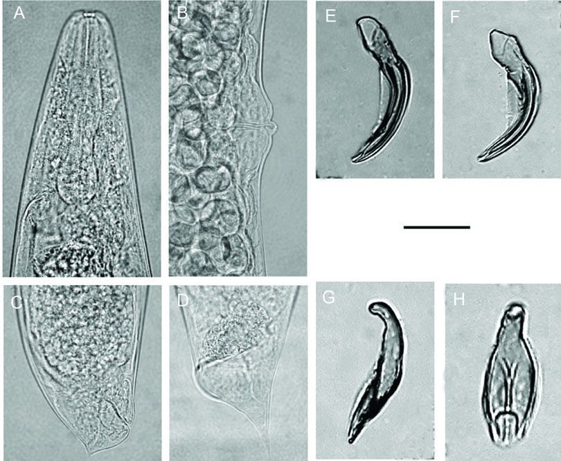
Body usually C-shaped on heat relaxation, cuticle smooth or
with faint annules. Lateral fields and phasmids absent. Head bluntly rounded,
continuous with body contour. Six labial, four cephalic papillae and two
amphids conspicuous. Stoma shallow. Cheilorhabdions well sclerotized but small.
Prorhabdions distinct. Pharynx muscular with cylindrical procorpus, metacarpus
swollen with a diameter similar to that of the basal bulb, isthmus distinct.
Excretory pore located anterior to nerve ring, near mid-pharynx. Gonads
amphidelphic, reflexed. Vulva a transverse slit, symmetrical and slightly
protruding from body surface. Double-flapped epiptygma present. (Fig. 3) Vagina short and muscular (Figs. 1B and 2B). The anal portion slightly
swelling. Tail conoid with tail length shorter than anal body diameter. A short,
projection-like mucron was observed on tail tip for about 80% of specimen
examined.
Female,
second-generation
Similar
to first generation female but smaller. Vulva slightly protruding from body surface.
Epiptygma present. Tail taperring to a pointy end, longer than anal body width.
Ventral postanal swelling present .
Infective juvenile
Body
elongate and ensheathed in second-stage cuticle right after harvesting, but
many of them will lose their sheath in storage or in soil. Head round
anteriorly and continuous with body contour. Labial region smooth. Four
cephalic papillae distinct. Amphids slit-shaped but not prominent. Cuticle
marked with transverse striations. pharynx long and thin. Excretory pore
located anterior to nerve ring (Fig. 3B).
Lateral field begins anteriorly with one line followed by two additional lines
to form two ridges. At about the level of excretory pore, each of the two
ridges separated into three, which makes the number of ridges increases to six
(Fig. 1J). The six-ridge pattern extents
posteriorly to the level of anus. After that, the ridges begin to merge, reduce
to two and then disappear. The six ridges are evenly distributed at least at
the middle body portion (Figs. 1K
and L). Thus, the formula of the lateral field is 2, 6, 2. Phasmid not
prominent. Tail attenuate and tapering gradually with constriction at the level
of hyaline portion. The hyaline portion accounted for about 54 % of tail
length.
Diagnotis characters
Steinernema
akhursti is
characterized morphologically by the combination of the features of various
developmental stages of the nematode. For the first-generation males, the new
species can be recognized by spicule length 90 μm; spicule tip blunt with
an aperture on the ventral side; gubernaculum with along and needle-shaped
cuneus; and tail conoid with a prominent mucron on the tip and a concave on
ventral side. The second generation males are characterized by a long and thin
mucron on the tail tip. For infective juvenile, the combination of the
following characters: body length (812 μ m), distance from anterior end to
excretory pore (59 μm), tail length (73 μm), E% (77), lateral field
with six evenly distributed and identical ridges at the middle body portion,
and tail with long and slightly constrict hyaline portion can be used to
differentiate the new species from other nematodes. For female, S. akhursti is
recognized by a conoid tail with a short mucron and slightly swelling anal
portion, and a slightly protruding and symmetrical vulva with conspicuous
double-flapped epiptygma. The new species is characterized genetically by its
unique sequences of both the 28S rDNA D3 domain and ITS regions, and it can be
separated from its closely related species S. feltiae and S.
oregonense by cross-breeding tests.
The
species is characterized genetically by sequence length of ITS regions and
partial 28S rDNA, 973 bp (246A, 234G, 304T, and 189C) (accession # DQ375757),
and 459 bp (108A,148G,
112T, and 91C) (accession # AY177188), respectively, and pair wise distances of these sequences
(Qiu et al. 2005).
Relationships
Both
molecular (Figs. 4
and 6) and
morphometric data showed that S. akhursti belongs to the S. feltiae group
and is closely related to S. feltiae, S. oregonense, Steinernema
kraussei (Steiner, 1923) Travassos,1927, S. weiseri, Steinernema sangi Phan,
Nguyen &Moens, 2001 , Steinernema thanhi Phan, Nguyen &
Moens,2001, and Steinernema monticolum Stock, Choo & Kaya,1997. The
shape of spicule and gubernaculum of the first-generation male, the tail and
lateral field morphology of infective juvenile, the tail and vulva shape of the
first-generation females, and the tail of the second-generation males (with a
long mucron) can be used to distinguish S. khursti to others. For
example, based on the shape of the first-generation male spicule, S.
akhursti (with well developed velum and an apertures on ventral side of the
tip, Fig. 1) can be easily separated from S.
feltiae and S. oregonense(without velum and apertures), S. sangi (head
length shorter than width) and S. thanhi (velum much thinner or
indistinct). For lateral field morphology, S. akhursti has six evenly
distributed ridges at mid body area while S. feltiae possesses eight
ridges with submarginal pair indistinct).
Type host and locality
The
type host of this nematode in nature is unknown as it was recovered from soil
using Galleria larvae as bait. The new species was recovered from many
soil samples collected from various localities in temperate areas of Yunnan
province during the surveys. An isolate(YNb112) was used as type specimens for
description. Itwas recovered from a soil sample collected from grassland in
Jiantang town, Zhongdian county, Diqing district,Yunnan province, China.
Type material
Holotype
(male), allotype female, paratype deposited in the State Key Lab for
Biocontrol, College of Life Sciences, Zhongshan University,Guangzhou 510275,
China. Some paratype specimen deposited
in UC Davis Nematode collection.
Distribution
About
30 isolates of this species were obtained from various localities in Yunnan
province. S. akhursti isolates were recovered from places with mean
annual temperature between 8 C (Zhongdian) and 16 C (Kunming, Dali and Qujing).
No S. akhursti was found in warm areas, such as Jinghong and Luxi
districts where extensive entomopathogenic nematode surveys have been carried
out (Qiu et al, 2005).
Symbiotic bacteria
The
symbiotic bacterium of S. akhursti has been isolated, purified, and
preserved in liquid nitrogen. The bacteria produced a red pigment on an
artificial medium containing corn flour, egg, and oil initially (Bedding, 1998) and then the color of the
medium gradually turned dark brown when nematodes proliferated. This feature of
pigment production was observed from all S. akhursti isolates, it is
unique for Steinernema species observed so far. Preliminary study
indicated that the symbiotic bacteria of S. akhursti possessed many
characters of Xenorhabdus species, such as phase variation, negative to
peroxide etc. A phylogenetic tree based on the 16S rDNA sequence showed that it
formed a monophyletic clade with, but can be clearly distinguished from, Xenorhabdus
nematophila and Xenorhabdus japonica (Qiu, et al. 2005).
Etymology:
This species is named after R. J. Akhurst from the Australian Center
for International Agricultural Research (ACIAR).
Reference
Qiu, L, Hu, X.., Zhou, Y., Mei, S., Nguyen K. B. & Pang, Y. (2005). Steinernema akhursti sp. n. (Nematoda: Steinernematidae) from Yunan, China. Journal of Invertebrate Pathology 90, 151-160.