Steinernema diaprepesi
Nguyen & Duncan, 2002
Steinernema diaprepesi
Nguyen & Duncan, 2002
Female face view Epiptygma Spicule
DESCRIPTION
Males, first generation:
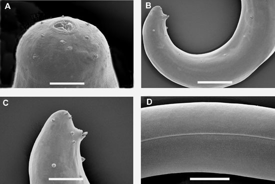
(SEM1) Body curved
ventrally posteriorly, C-shaped when heat-killed. Lateral field present
in mid-body with one narrow ridge (SEM1D).
Head rounded, usually slightly swollen (SEM1A).
Anterior end with a perioral disc around stoma; six
labial papillae, two amphids (usually covered with exudate) and
four cephalic papillae (SEM1A).
Stoma shallow, cheilorhabdions as small and sclerotized structures at anterior
end (Fig1A), sometimes
indistinct. Excretory pore near nerve ring, located mostly anterior
to basal bulb. Esophagus with cylindrical procorpus, metacorpus absent
or slightly swollen, isthmus present, nerve ring around isthmus, basal
bulb distinct. Esophago-intestinal valve present, usually weak. Gonad
monorchic, reflexed. Distance from base of esophagus to anterior
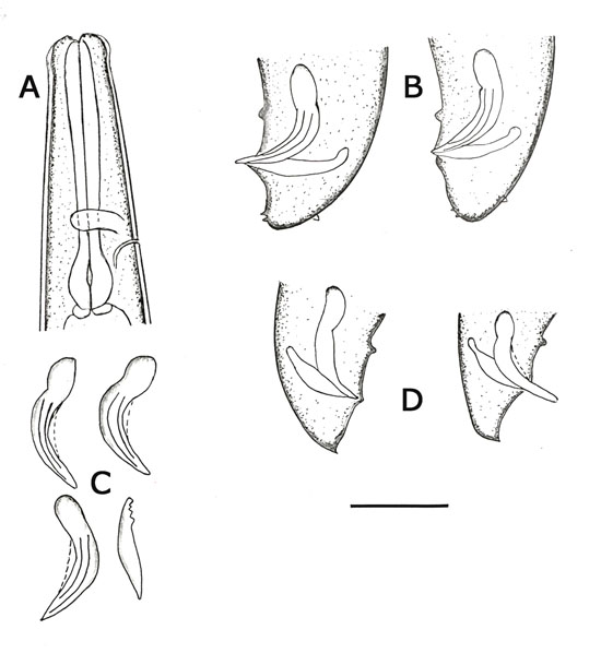
end of testis variable. Spicules (SEM2)
paired, brown in color. Head of spicules elongate, the ratio length/width
from 1.4-2.0 (averaging 1.7 ± 0.2) in some specimens (4/20), twice
as long as wide; shaft very short or absent; blade thick, tapering
gradually posteriorly, blade terminus blunt with a longitudinal depression
slit in ventral side (SEM2C,D);
velum present. Each spicule with two internal ribs. Gubernaculum
boat-shaped in lateral view, anterior part usually with one or two ventral
projections (SEM2C,
2G). Eleven pairs [occasionally twelve (SEM1B)]
and one single precloacal genital papillae. Tail conoid; tail terminus
without mucron (SEM11B,
2B).
General morphological characteristics of males collected from
Diaprepes
abreviatus appear similar to those collected from
Galleria
mellonella, but they differ morphometrically. Most measurements
of first-generation males from D. abreviatus are shorter than
those collected from G. mellonella.
Paratype measurements: (Male, first generation,
collected from Galleria mellonella n=20). L= 1735 (1506-2078)
um; W=113 (90-145) um; EP= 115(100-130)um; ES=150 (136-162) um; anal body
width=42 (36-50) um; tail = 25 (20-32) um; spicule length along the arc
= 79 (71-90) um; gubernaculum length = 54 (45-61) um; D%= 80
(68-86); SW = 1.8 (1.5-2.0), GS =0.69 (0.59-0.79).
Males, second
generation: Second-generation male similar to
that of the first generation except body, spicule and gubernaculum shorter
and thinner, excretory pore much more anterior, and isthmus more distinct.
Gubernaculum short. Mucron on tail terminus sometimes present (Fig1D).
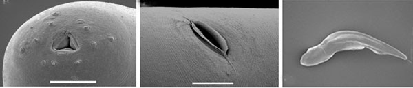
Females, first
generation: (SEM3)
Body
cuticle smooth or with faint annules. Lateral fields and phasmids absent.
Head rounded, continuous with body; six labial papillae, four cephalic
papillae (SEM3A, B).
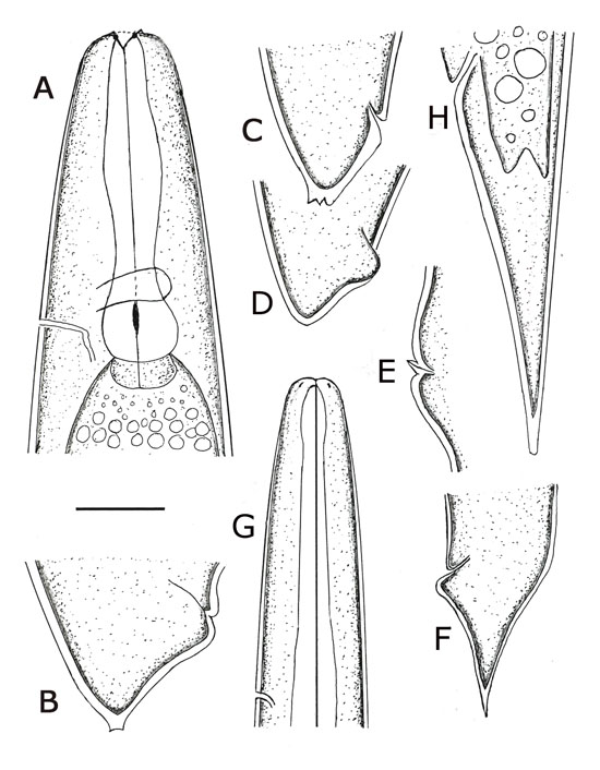
Lips indistinct. Amphids present. Stoma shallow, subtriangular anteriorly;
triradiate internally (SEM34A,
B). Cheilorhabdions (Fig2A),
well sclerotized but small. A smaller sclerotized structure posterior
to cheilorhabdions (presumably the prorhabdions), observed in other species,
indistinct in this species. Esophagus with procorpus cylindrical,
muscular; metacorpus swollen; isthmus distinct; basal bulb valvate (Fig2A)
as in other steinernematids. Nerve ring surrounds isthmus, just anterior
to basal bulb. Esophago-intestinal valve present. Excretory
pore located just anterior to, or at the middle of basal bulb. Gonads amphidelphic,
reflexed, often containing many eggs. Vulva, a transverse slit; protruding
or not; low double-flapped epiptygma present (SEM3E,
F). Vagina sclerotized, short. Body width greater anterior
to vulva than posterior to vulva. Tail shape variable (Fig. 5B, C),
ventral postanal swelling present, tail shorter than anal body width.
Most females with five papillae-like structures on tail tip (SEM3C,
D). The size of these structures is variable depending on age of females,
usually longer in young females (Fig. 5C), becoming smaller and short as
the female bodies enlarge, disappearing in fully mature females (Fig2D).
Females, second generation:
Similar to first generation female but smaller (length = 2385 um, width
= 149 um compared to 6512 um and 264 um, for first-generation female).
Vulva less protruding, epiptygma (Fig2D)
usually more prominent than that of first generation females. Tail, tapering
to a pointy end, longer than anal body width; ventral postanal swelling
present (Fig2E).
Infective juveniles:
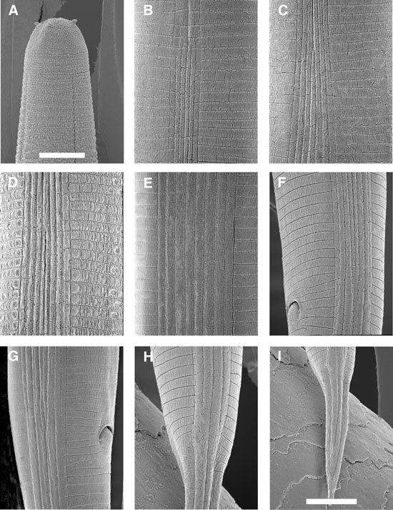
(SEM4)Body elongate.
Sheath (second-stage cuticle) present immediately after harvesting, but
many IJ will lose their sheath in storage. Labial region smooth,
continuous, rounded anteriorly (SEM4A).
Labial papillae not seen; four cephalic papillae prominent. Amphids
slit-shaped but not prominent. Cuticle marked with prominent transverse
striations. Lateral field begins anteriorly with one line. Two additional
lines appear at annules 14-16 to form two ridges (SEM4B).
Near excretory pore level, the number of ridges in lateral fields increases
from two to
six . A new central ridge appears more posteriorly, about a body
width posterior to excretory pore, making a total of seven ridges in the
lateral field (Fig. 6C). Near the end of esophagus, the central ridge divides
into two, making a total of eight ridges, the maximum number in the lateral
field (SEM4D, E). The
portion with eight ridges is the longest part (compared to portions with
2, 6, 7, 4 ridges) of the lateral field. At the level of the anus the two
marginal and two central ridges disappear (SEM4F,
G), only four ridges remain in the lateral field. At about mid-tail
the four ridges in the lateral field become two ridges (SEM4H).
Near tail terminus, the two marginal lines in the lateral field converge
(SEM4I), and the central
line disappears before reaching the end of the lateral field.
Esophagus with thin corpus (Fig2G),
basal bulb more or less elongate with visible valve. Tail attenuate, tapering
gradually (Fig2H).
Hyaline portion occupies about 57% (50-63) of tail length, sometimes, up
to 70% (Fig. 5G).
The morphometrics of infective juveniles reared from G. mellonella
and from D. abbreviatus are different.
Measurements (n=20, all measurements are in micrometers): L
= 1002 (880-1133) ; W = 34 (30-42); EP = 74 (66-83); ES = 138 (111-152);
ABW = 23 (21-27); tail = 83 (65-91); a = 30 (23-35); b = 7.3 (6.5-8.3);
c = 12.1 (10.4-13.2); hyaline part/tail = 57 (50-63); E% = 90 (78-114);
D% = 54 (30-70).
DIANOSIS:
Males: Spicule averaging 79 (71-90) um; D% about 80;
the ratio SW about 1.8. Lateral field with one narrow ridge. Females:
Vulva with short, low double-flapped epiptygma; tail terminus usually with
5 papillae-like structures. Infective juveniles: Body
length averaging 1002 (880-1133) um, EP = 74 (66-83) um; tail length =
83 (65-91) um, and E% = 89.6 (78-114). Lateral field pattern variable,
the formula for the arrangement of ridges from head to tail is: 2, 6, 7,
8, 4, 2 (SEM4).
The portion with 8 ridges is the longest. The new species is characterized
genetically by sequence lengths of ITS region (808 bp), ITS1 (301 bp),
ITS2 (313 bp), and composition of its sequence (primers AB28 and TW81were
used for PCR).
BIOLOGY:
Steinernema diaprepesi
was isolated from larvae of D. abbreviatus that were buried in cages
beneath citrus trees . During two years of the experiment, indigenous
populations of S. diaprepesi . infected and killed 13-50% of the
buried insects within seven days, with higher rates of parasitism during
the summer months compared to spring and autumn months. The nematode
was isolated with much lower frequency from the rhizosphere of native plants
growing between citrus tree rows than from the rhizosphere of citrus trees.
The topotype locality is an irrigated, commercial citrus orchard c. 6 k
east of Bartow, and immediately southeast of the intersection of Cowpen
Road and 80-Foot Road. Mean monthly temperatures range from
16.1-27.9 o C and cumulative annual precipitation averages 1364 mm with
most rainfall occurring May-September and frequent periods of drought from
mid-autumn to late-spring. Soil texture is astatula sand (97:1:2,
sand:silt:clay). S. diaprepesi has been isolated from
other orchards located on the central ridge of Florida which is characterized
by deep sandy soils. Attempts to isolate the nematode in heavier-textured
soils off of the central ridge have been unsuccessful; however, no comprehensive
survey has yet been conducted.
Reference
Nguyen, K.B. and L. W. Duncan:Steinernema
diprepesi n. sp. (Rhabditida: Steinernematidae), a parasiteof the citruc
weevil Diaprepes abbreviatus (L)
(Coleoptera: Curculionidae). Journal of Nematology
34:159-170.